The Laboratory of Laser Molecular Spectroscopy consists of seven laboratories:
- Raman Spectroscopy Laboratory
- Raman Imaging Laboratory
- Infrared Spectroscopy and Imaging Laboratory
- AFM Imaging Laboratory
- NanoIR-AFM Laboratory
- Femtosecond Spectroscopy Laboratory
- Molecular and Cellular Biology Laboratory
Raman Imaging Laboratory
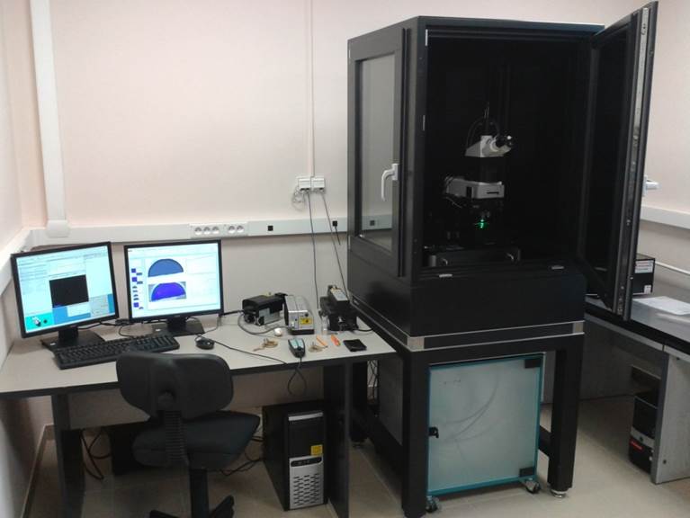
The Confocal Raman/AFM/SNOM/TERS Microscope (WITec alpha 300RSA+) enables the acquisition of 2D and 3D Raman maps, Raman spectra, SNOM imaging, and AFM maps. Thanks to its unique design, Raman/AFM/SNOM analysis can be performed on the same sample location without changing its position.
The microscope is equipped with lasers at wavelengths of 355, 532, and 785 nm, as well as two monochromators and diffraction gratings with 1200, 2400, and 3600 lines/mm. The objective set includes 20x, 40x, 60x, 100x objectives, and a 40x objective for measurements in liquids.
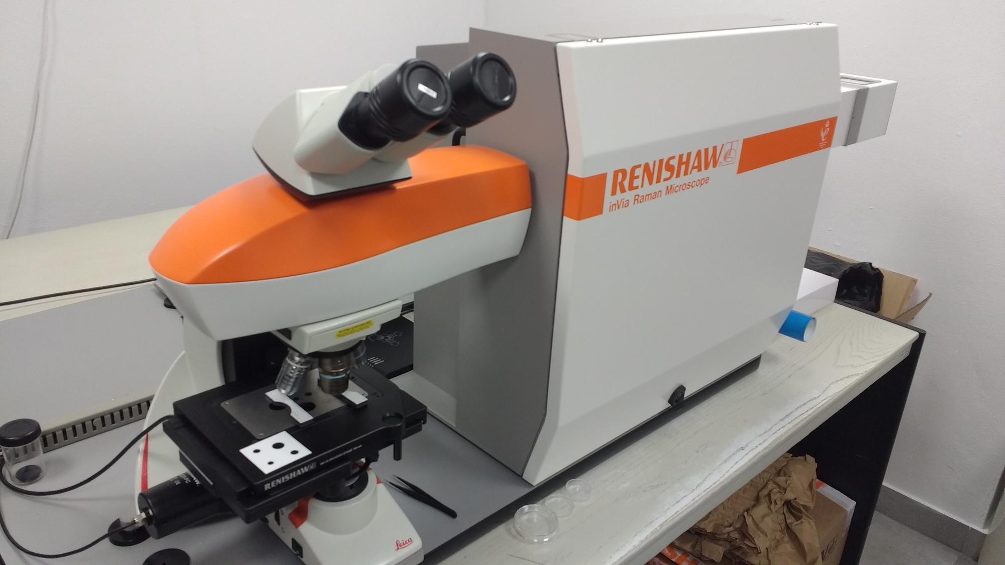
The Confocal Raman Microscope (Renishaw) allows for the acquisition of Raman spectra and Raman maps. The system is equipped with two excitation lines at 532 and 633 nm, and it includes a set of objectives with magnifications of 10x, 40x, 50x, and 100x, as well as diffraction gratings with 600 and 1800 lines/mm.
Infrared Spectroscopy and Imaging Laboratory
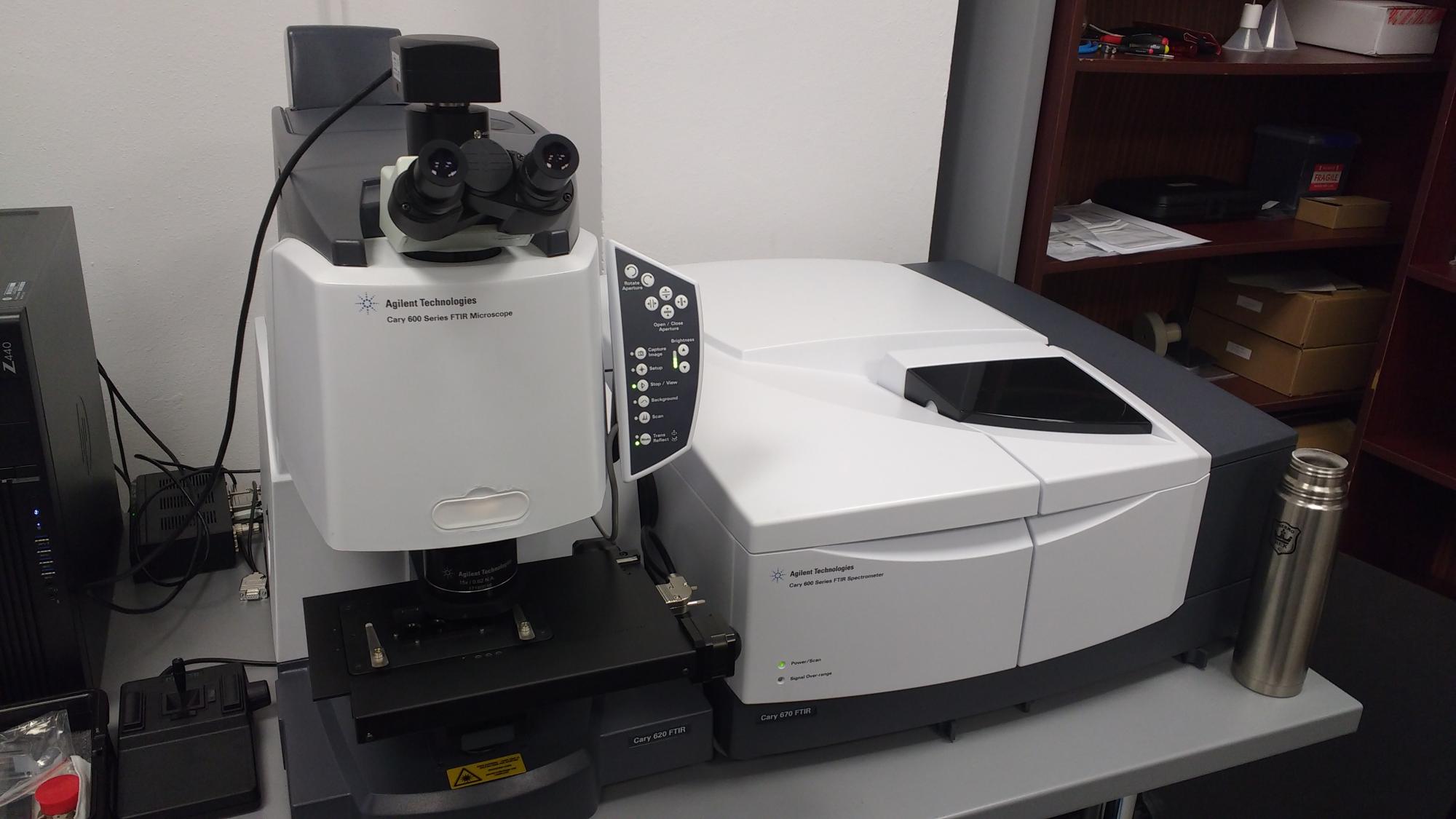
The Agilent Technologies Cary 600 Series FTIR Microscope is an IR spectrometer coupled with a microscope for recording IR spectra and IR imaging. It consists of a spectrometer equipped with an interferometer mounted on an air-bearing bench. The spectral range of the device is 350-9000 cm-1 with a spectral resolution of 0.075 cm-1. It is equipped with a Peltier-cooled DTGS detector with a signal-to-noise ratio of 50,000:1, and it offers a high-speed scan function of 110 spectra per second at a resolution of 16 cm-1. Additionally, it features a dual analog-to-digital converter.
The microscope allows measurements in transmission, reflection, and ATR modes, and it includes a set of objectives: 15x for the IR range and 4x for the visible range. It has a highly sensitive 15x condenser and is equipped with detectors, including a single-element MCT detector, an FPA matrix detector with a 64x64 pixel array, and a trinocular head with a high-resolution 14 Mpixel camera.
AFM Imaging Laboratory
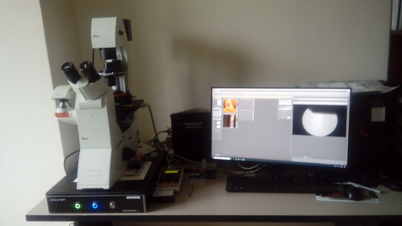
The AFM Microscope (Pik Instruments) enables the acquisition of AFM maps and the analysis of nanomechanical parameters. The microscope is equipped with a controller with a scanning range of 100 x 100 µm in the X and Y axes and 15 µm in the Z axis, with positioning resolution in the XY axes of 6 pm and in the Z axis of 0.9 pm. The microscope has an inverted configuration. The system allows measurements in both air and liquids, in contact and tapping modes. The software allows for the generation of maps of mechanical properties, including topography, stiffness, adhesion, and corresponding histograms.
Femtosecond Spectroscopy Laboratory
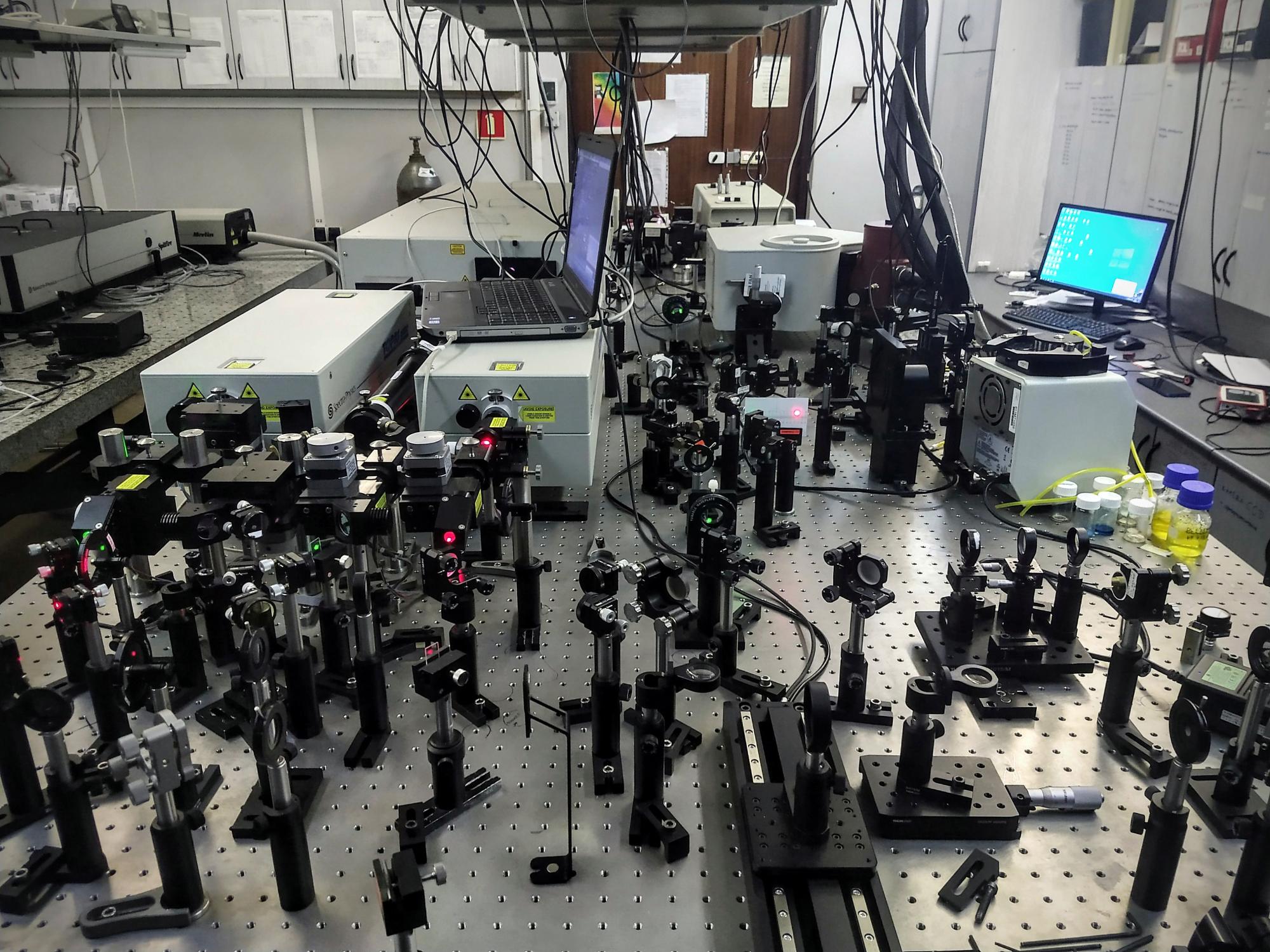
The femtosecond system enables the study of ultrafast photochemical and photophysical processes. The system consists of the following components:
- Femtosecond Titanium-Sapphire laser Ti+3Al2O3 (Tsunami, Spectra Physics), pulse duration: 100 fs, wavelength 798 nm, repetition rate 82 MHz - pumping laser for the femtosecond laser: solid-state laser (Nd:YVO4) (Millennia-Pro, Spectra Physics), 532 nm, 5 W - regenerative amplifier (Spitfire Ace, Spectra Physics), wavelength 800 nm, repetition rate 1 kHz, average output beam power 4 W, pulse duration: 100 fs.
- Pumping laser for the regenerative amplifier: Q-switched solid-state Nd:YLF laser (Empower 30, Spectra Physics), wavelength 527 nm, repetition rate 1 kHz, minimum output beam power 20 W."
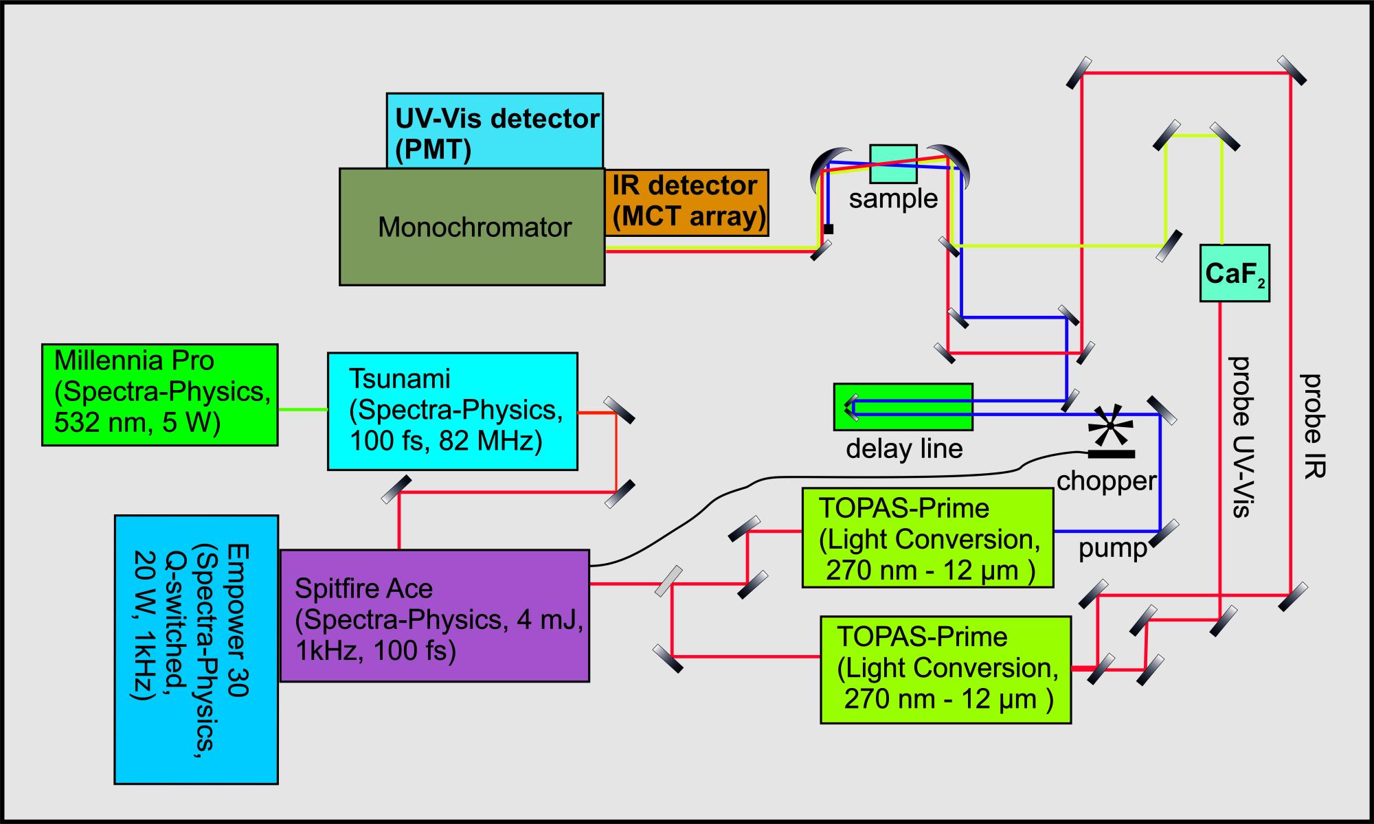
- Two Optical Parametric Amplifiers (OPA) allowing for spectral tuning of femtosecond pulses in the range of 290-11000 nm (TOPAS, Spectra Physics).
- Phase-locked amplifier (SRS 830, Stanford Research Systems).
- HgCdTe detector (MCT): a 2x64 element array with data acquisition system (FPAS-0144, Infrared Associates) for femtosecond transient absorption measurements in the IR range.
- Monochromator (iHR320, HORIBA Scientific).
- Photomultiplier Tube (PMTSS, Thorlabs) for highly sensitive transient absorption measurements in the spectral range of 185-900 nm.
- Chopper (Model 3502, Newport)
Raman Spectroscopy Laboratory
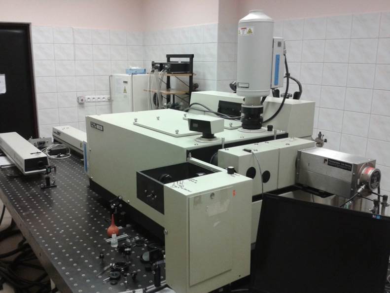
The Raman spectrometer allows for the registration of Raman spectra.
The Raman spectrometer U 1000 (Jobin Yvon) comprises the following components:
-
Argon ion laser (Stabilite 2017-Spectra Physics), wavelength 454-514 nm, CW laser, output beam power 100 mW-2 W depending on the wavelength of the emitted beam (488 nm – 1.5 W; 514 nm – 2 W).
-
CCD camera (Horiba, Jobin-Yvon, CCD - 1024x256 - open -3LD).
-
Cryostat (Oxford Research Limited).
Molecular and Cellular Biology Laboratory
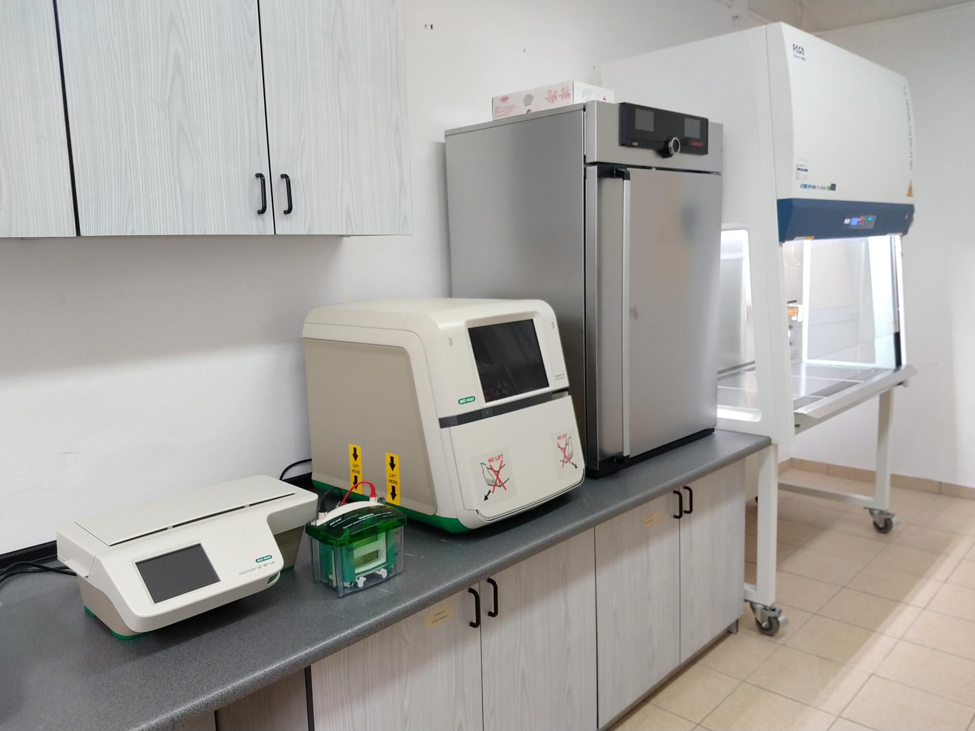
The set for the analysis of fluorescence and chemiluminescence in biological systems allows for the visualization of proteins and antibodies. It is equipped with kits for protein isolation and staining based on Western blot analysis, which includes sample preparation with the analyzed protein mixture, electrophoresis, transferring separated proteins from the gel to a membrane, incubation with appropriate antibodies, and detection of the desired protein.
The system includes:
- Vertical electrophoresis unit
- Two-level laboratory shaker
- Rapid protein transfer apparatus
- Universal power supply for use in the electrophoresis process
The device enables visualization, recording, and analysis of fluorescently, colorimetrically, and chemiluminescently labeled samples using gel-free technology.
- A system designed for isoelectric focusing of proteins (IEF) in strips with immobilized pH gradient (IPG) for the first dimension of two-dimensional (2-D) electrophoresis.
- Electrophoresis kit with IEF-focused IPG strips to obtain a 'two-dimensional protein map' in 2D electrophoresis.
- Centrifuge with accessories.
- Class II laminar flow cabinet for safety.
- Cell culture incubator with CO2 level control.
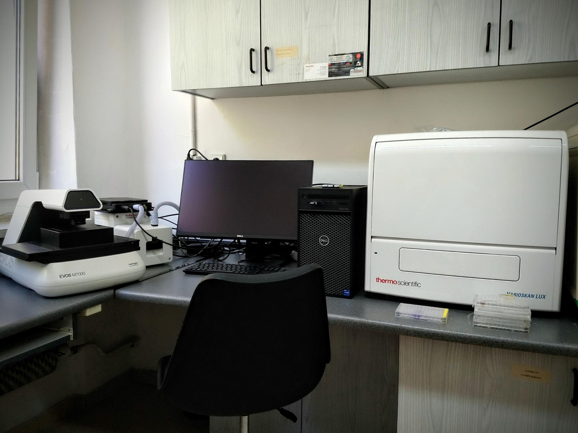
The cell viability analysis kit, based on absorbance and fluorescence measurements, is equipped with an automated inverted fluorescence microscope for brightfield, fluorescence, and phase-contrast cellular imaging. It is also equipped with a multi-detector microplate reader capable of detecting fluorescence intensity, fluorescence polarization, luminescence, and absorbance. Detection is possible for both 96 and 384-well plates. The kit ensures complete control over imaging conditions, including temperature, humidity, and atmospheric composition (CO2/O2), allowing for measurements under cell culture conditions. It includes a fully isolated imaging chamber that can be heated to 60°C. The system also features an external peristaltic pump module for the direct dispensing of various reagents into the designated microplate well
NanoIR-AFM Laboratory
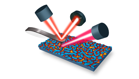
The nanoscale IR-AFM imaging system enables simultaneous measurements of IR absorption (IR spectra), sample topography, and nanomechanical properties (AFM) based on the photothermally induced expansion effect, with an extreme spatial resolution (<10 nm) based on measurements of the change in frequency and amplitude of the AFM probe.
The system comprises:
- An infrared microscope enabling measurements with a nanometric spatial resolution of no less than 10 nm.
- An infrared radiation source in the form of a tunable QCL (Quantum Cascade Laser) pulse laser with a wavelength range of at least 800-1800 cm-1, allowing for the recording of a full IR spectrum in this range in a maximum of 10 seconds, with a spectral resolution of at least 1 cm-1.
- The system allows for operation in a resonant enhancement mode, where the repetition frequency of the IR source pulses can be matched to the frequency
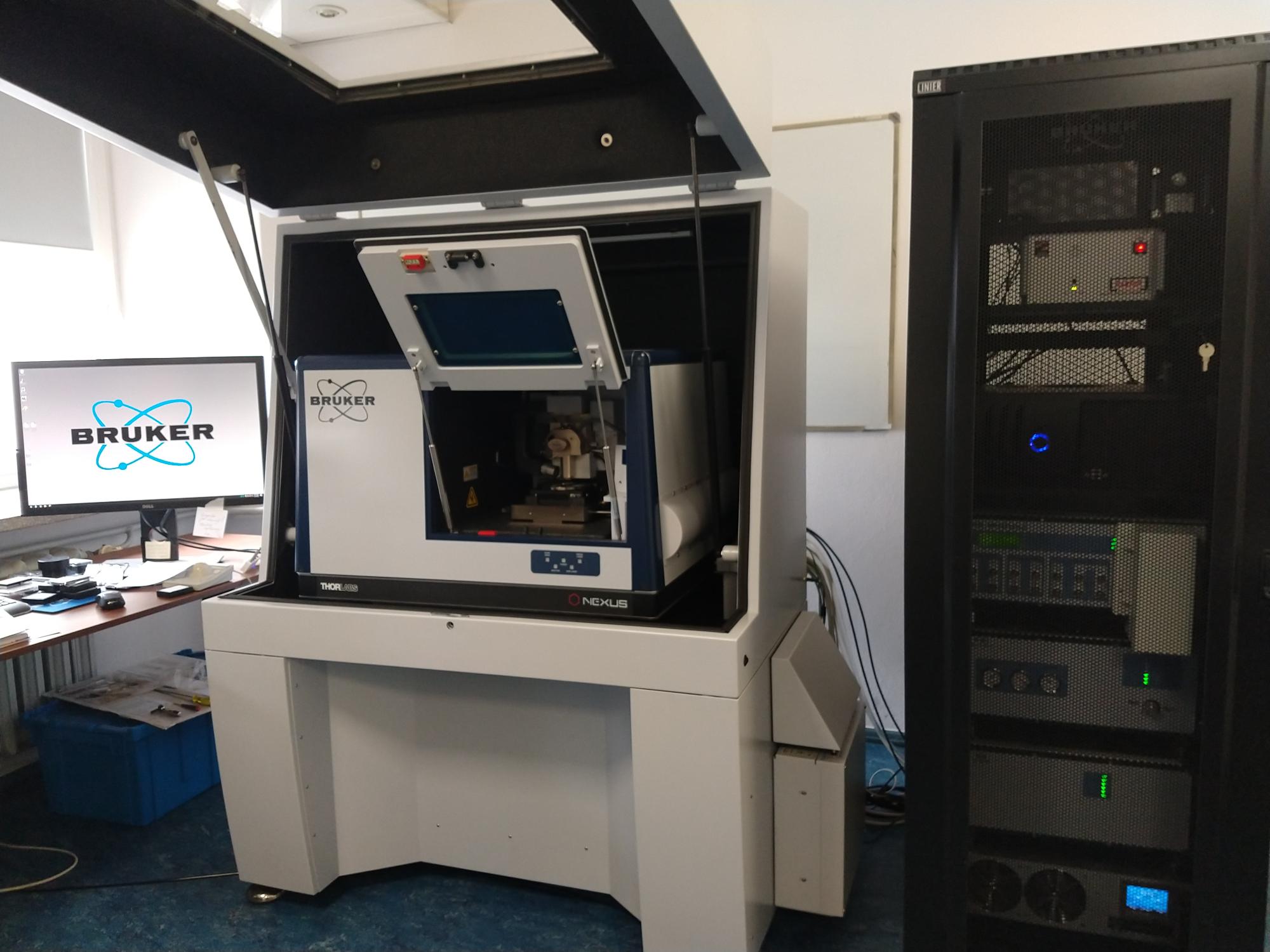
Characteristics of the nanoIR-AFM system
• Extreme spatial resolution <10 nm
• Rapid chemical imaging
• Nanoscale FTIR
• Highest nano-IR performance utilizing AFM-IR and FASTspectra modules